Written by Nicolai Garcia
On August 28, we went to the Conservation Center (Institute of Fine Arts, New York University) housed in a historic town house on the Upper East Side to x-radiograph some electronic objects in Cooper Hewitt’s collection. The x-ray room, which doubles as a photography studio, is entirely black from floor to ceiling. Behind a door—reminiscent of those in sci-fi nuclear thrillers—sits the x-ray machine, black and medical.
Over the course of the morning, we unboxed an array of objects from Cooper Hewitt’s collection dating from the early 1980s to the present. We laid them onto an x-ray-sensitive sheet (which was wrapped in vinyl as to not expose the sheet to light) and then exposed them to the x-ray beam for a short amount of time (like a photographic exposure). Once the machine was done, the lights in the studio were turned off while we loaded the x-ray plate (like a photographic negative) onto a scanner.
We sat in anticipation while the image rendered, layer by layer, giving us a glimpse into each object’s concealed interior. In some cases, the rendering was over-exposed, meaning we had to do another round, with a lower voltage or shorter exposure. Andy Wolf, a current Conservation Center graduate student who assisted in the examination, compared adjusting the voltage and exposure of an x-ray beam to adjusting the strength and open area of a faucet; both control how much water comes out, but in different ways.
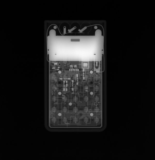
X-radiograph of a Braun calculator (2015-5-4) in the Cooper Hewitt collection, taken at 25 kV, 5mA with a 30s exposure.
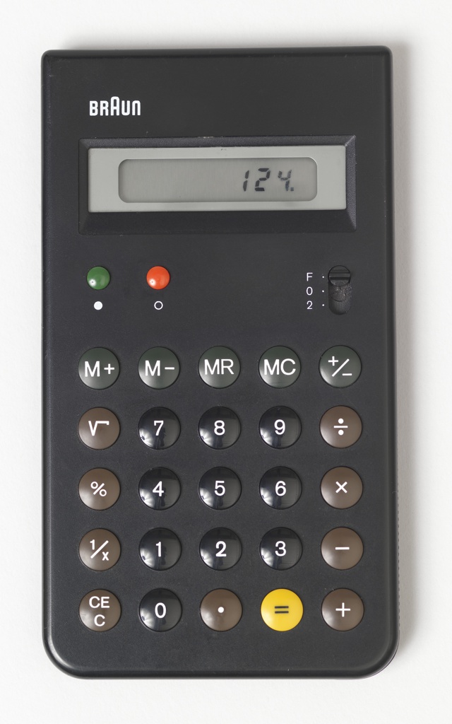
Exterior of ET55 Calculator, 1980; Designed by Dieter Rams (German, b. 1932) and Dietrich Lubs (German, b. 1938); Manufactured by Braun AG (Frankfurt, Germany); Molded ABS plastic, electronic components; H x W x D: 13.6 × 7.6 × 1 cm (5 3/8 in. × 3 in. × 3/8 in.); Gift of George R. Kravis II, 2015-5-4
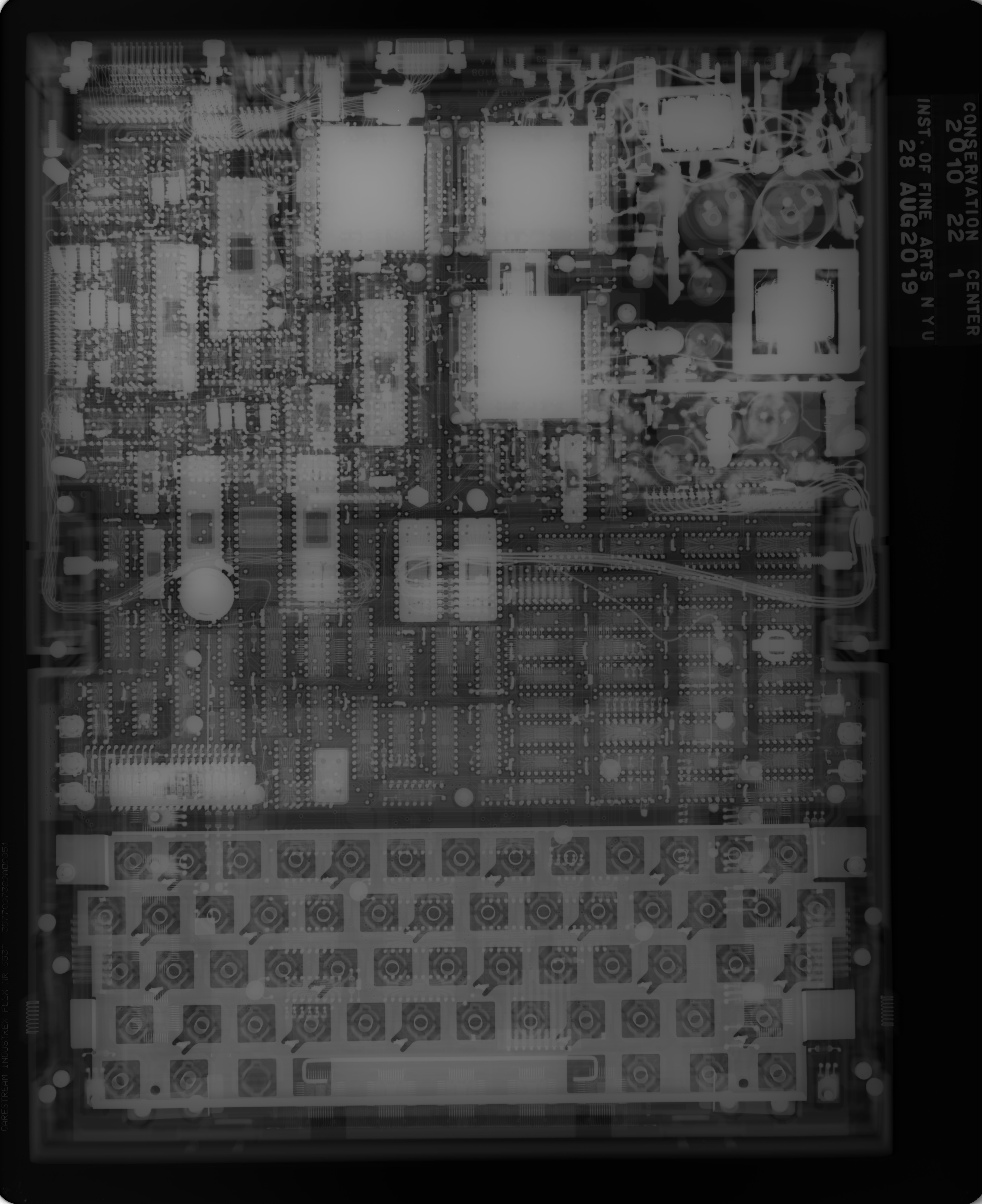
Digital x-radiograph of a GRiD Compass laptop (2010-22-1) in the Cooper Hewitt collection, taken at 70 kV, 5mA, with a 60s exposure.
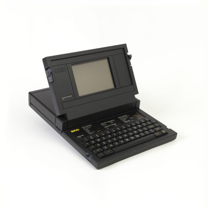
Exterior of GRiD Compass Laptop Computer Prototype, 1981; Designed by Bill Moggridge (English, 1943–2012); Manufactured by GRiD Systems Corps (Mountain View, California, USA); Die-cast magnesium, injection-molded plastic; H x W x D: 25.4 x 29.2 x 37.9 cm (10 in. x 11 1/2 in. x 14 15/16 in.); Gift of Bill Moggridge, 2010-22-1
In the case of the objects we imaged, adjusting the voltage versus exposure depended on the object’s material. The voltage, measured in kilovolts (1,000 volts), is typically increased for materials made with elements with a higher atomic number. Steel, which is an iron-based metal with just a little bit of carbon, has a lower density and therefore requires a lower x-ray voltage compared to platinum, which has an atomic number close to lead, meaning it requires a high voltage in order for the x-ray beam to penetrate its surface. Lead is dense and heavy enough to be impenetrable to x-rays, and is used to line the x-ray room to protect those nearby from stray radiation. For reference, the voltage for a medical x-ray of a human being (carbon-based life-form!) hovers around 60 kv.
We repeated the cycle of imaging and adjusting for all the electronics objects (save for a platinum-encased cell phone, which our x-ray beam did not have high enough voltage to image), then took the images to analyze what we saw.
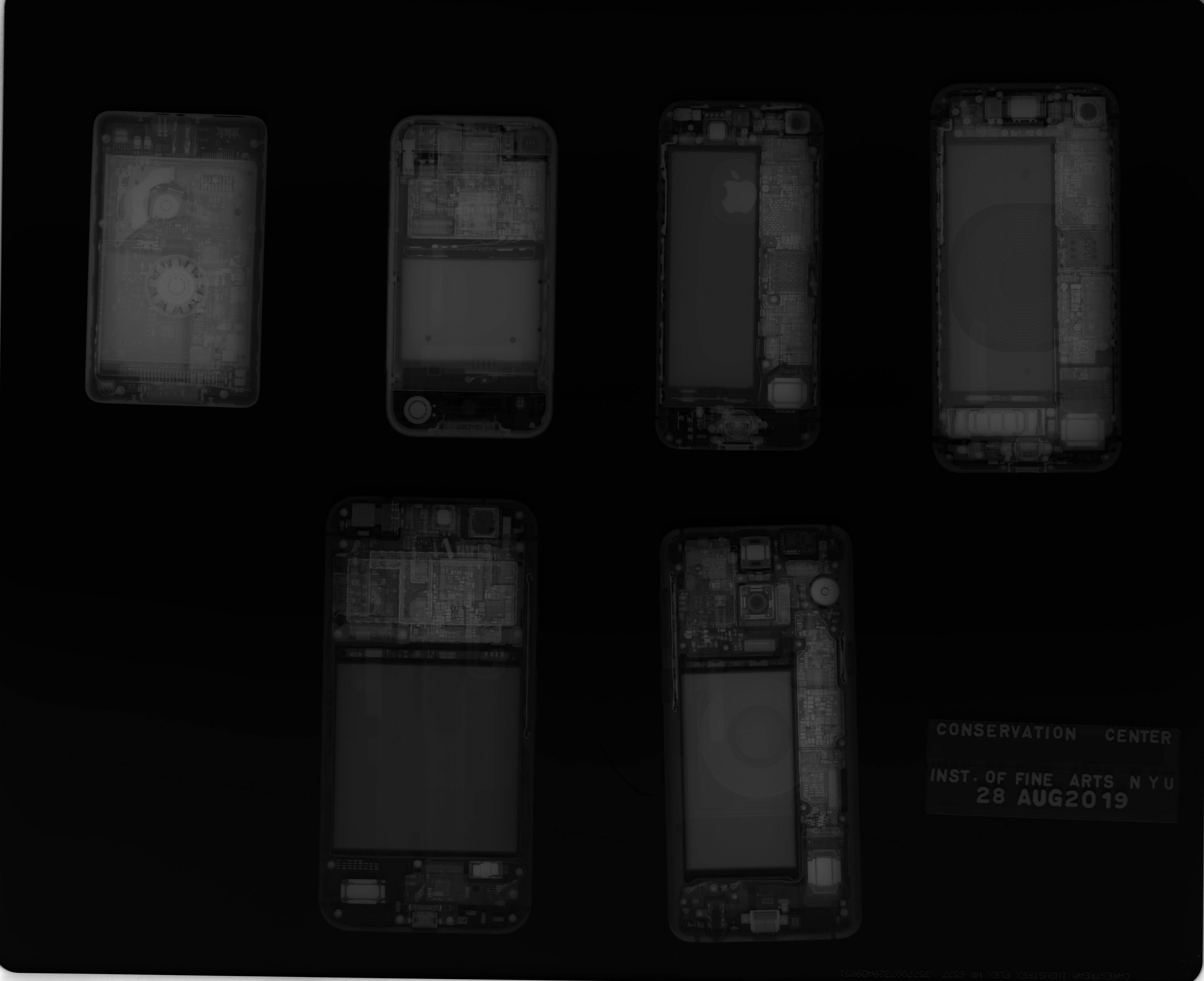
Digital X-radiograph of collections objects including a 2007 iPhone and a 2014 Pixel phone shown with contemporary cell phones (iPhone SE, iPhone 8, and Samsung Galaxy).
In the current age of technology, common devices, such as mobile phones and home assistants, are designed to hide their screws and other traces of how their components come together, making it hard to know what they look like under the hood. X-ray imaging these objects gives conservators a better idea of the structural makeup so that they understand what techniques must be applied to preserve them. In my experience seeing the x-ray of my iPhone 8 (far right), I felt more connected to it because I feel I saw who it really is inside.
Nicolai Garcia was the Digital Collection Intern at Cooper Hewitt during summer 2019. Nico is from Berkeley, California, and is a senior at Stanford University where he studies Computer Science and Art Practice.
Our thanks to the faculty and staff at the Conservation Center for allowing us to use their X-radiography equipment, and to Andy Wolf for his help!
One thought on “X-Ray Vision: Exposing 40 Years of Gadgets”
Marco Garcia on October 21, 2019 at 1:35 pm
Terrific article! Interesting to read how the X-ray machine can be adjusted for different materials by tuning the voltage and exposure.